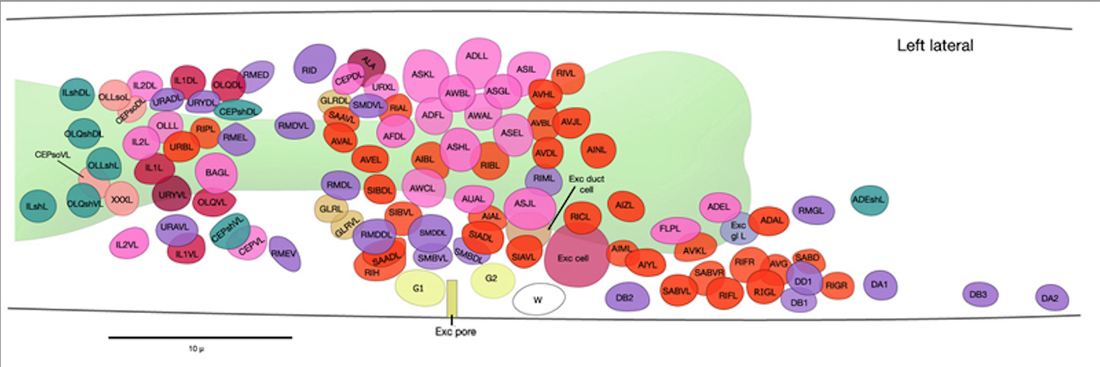
The excretory system removes nitrogenous waste in the form of ammonia through the body wall. Amino acids, peptides, amines, carbon dioxide, fatty acids, and urea, are also excreted by nematodes. The cells that make up the excretory system are: one pore cell, one canal cell, one duct cell, and a fused pair of gland cells.
- The excretory cell plays a osmotic/ionic regulation and waste elimination role.
- The excretory cell collects fluids and then empties through the excretory duct and pore.
- Materials are also secreted from large membrane-bound vesicles.
- The secretion from the excretory canal and gland cells pass through a cuticle lined duct under the pharynx, in the excretory duct cell.
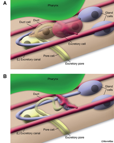
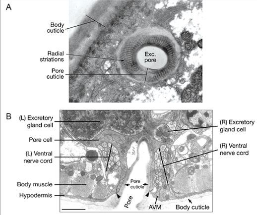
- The excretory cell, the gland cell, and the duct cell are all joined at the specialized intercellular junction called the secretory-excretory junction, where secretion from the glands and excretion pass through the duct to the outside of the body.
- In the bottom image, the excretory cell and duct cell are lifted to show the junction (pink jagged thing).
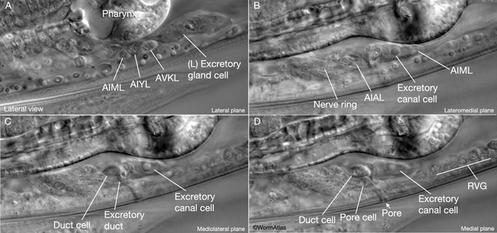
Different views of the excretory system: (Inside C. elegans)
Image A: Excretory gland cell, located on the ventral side between the intestine and the terminal bulb of the pharynx.
Image B: The excretory cell nucleus has a "fried egg" appearance with a large nucleolus.
Image C: The duct is anterior to the excretory cell.
Image D: The excretory pore cell is ventral to the duct cell. The duct passes thr
Excretory Canal Cell:
- The largest cell inside the nematode.
- Lies below the terminal bulb of the pharynx, next to the ventral epidermal ridge, acts as a connector between the right and left excretory canals.
- The two surfaces, basal and apical, join at the secretory-excretory junction.
- The canal stretches from the nose to the tail region.
- Run along the basolateral surface of the hypodermis.
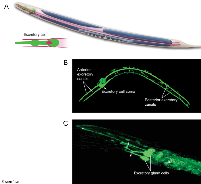
Excretory Gland Cell:
- Binucleate cell formed by the fusion of two identical cells.
- Separate cell bodies along the dorsal surface of the ventral nerve cord and fuses with the other side at the secretory-excretory junction. They fuse again to form a ring-like process, which projects to the nerve ring. The gland cells receives synaptic input from nerve ring neurons in this region.
- The cytoplasm contains a rough endoplasmic reticule, many mitochondria and ribosomes, Golgi complexes, and bunches of electron dense secretory granules. These granules are grouped around the cytoplasmic bridge region near the secretory membrane, which connects to the origin of the excretory duct.
Excretory Duct Cell:
- Cuticle lined channel that connect the excretory system to the outside of the body, via the excretory pore cell.
- It is located to the side of the excretory cell.
- Placement of the duct is based on the zinc-finger gene.
- Inside, the plasma membrane is invaginates (turns inside out), creating lamellar stacks, which increase the membrane's size. These stacks are similar to the hypodermis.
- Mutations can slow the growth of the duct cell, during embryogenesis, which can lead to death if the mutation is serious enough.
- Without drainage of the duct, excretory system, fluid accumulation can lead to death, because of the build-up of harmful waste.
Excretory Pore Cell:
- The pore cell is a specialized, transitional, epithelial cell.
- It closes off the end of the duct and forms junctions with the duct cell-pore cell junction. It also wraps itself around the duct.
- Similar with the duct cell, mutations can cause death of the organism due to build-up of hazardous waste.
- The duct cell and pore cell are responsible for creating a new cuticle after each molt.